1887

Graphical Abstract

 Graphical abstract 

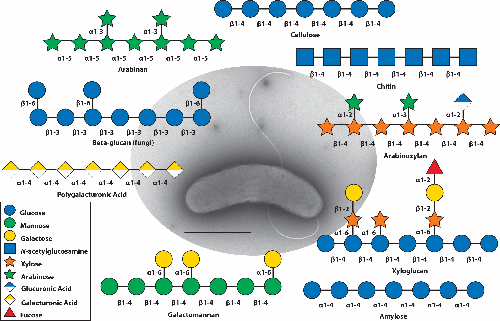
Transmission Election Microscopy (TEM) image of from a Hitachi HT7800 set to 100kV and stained with NanoW. The scalebar depicts 1µm. The polysaccharide diagrams shown are examples of substrates that the bacterium is adept at degrading. possesses an impressive array of carbohydrate active enzymes to cleave the glycosidic bonds of many plant, animal, and fungal polysaccharides.


Abstract

is a saprophytic bacterium proficient at environmental polysaccharide degradation for carbon and energy acquisition. Genetic, enzymatic, and structural characterization of carbohydrate active enzymes, specifically those that degrade plant and animal-derived polysaccharides, demonstrated that this bacterium is a carbohydrate-bioconversion specialist. Structural analyses of these enzymes identified highly specialized carbohydrate binding modules that facilitate activity. Steady progress has been made in developing genetic tools for to better understand the function and regulation of the polysaccharide-degrading enzymes it possesses, as well as to develop it as a biotechnology platform to produce renewable fuels and chemicals.

Funding
This study was supported by the:
  • National Institute of General Medical Sciences (Award R01GM147142-01)
    • Principle Award Recipient: JeffreyGardner
  • This is an open-access article distributed under the terms of the Creative Commons Attribution License.
Loading

Article metrics loading...

/content/journal/micro/10.1099/mic.0.001450
2024-04-03
2024-04-29
Loading full text...

Full text loading...

/deliver/fulltext/micro/170/3/mic001450.html?itemId=/content/journal/micro/10.1099/mic.0.001450&mimeType=html&fmt=ahah

References

  1. Ueda K, Ishikawa S, Itami T, Asai T. Studies on the aerobic mesophilic cellulose-decomposing bacteria. part 5-2. Taxonomical study on genus Pseudomonas. J Agric Chem Soc 1952; 26: [View Article]
    [Google Scholar]
  2. DeBoy RT, Mongodin EF, Fouts DE, Tailford LE, Khouri H et al. Insights into plant cell wall degradation from the genome sequence of the soil bacterium Cellvibrio japonicus. J Bacteriol 2008; 190:5455–5463 [View Article] [PubMed]
    [Google Scholar]
  3. Gardner JG. Polysaccharide degradation systems of the saprophytic bacterium Cellvibrio japonicus. World J Microbiol Biotechnol 2016; 32:121 [View Article] [PubMed]
    [Google Scholar]
  4. Gardner JG, Keating DH. Genetic and functional genomic approaches for the study of plant cell wall degradation in Cellvibrio japonicus. Meth Enzymol 2012; 510:331–347
    [Google Scholar]
  5. Hazlewood GP, Gilbert HJ. Structure and function analysis of Pseudomonas plant cell wall hydrolases. Prog Nucleic Acid Res Mol Biol 1998; 61:211–241 [View Article] [PubMed]
    [Google Scholar]
  6. Forsberg Z, Nelson CE, Dalhus B, Mekasha S, Loose JSM et al. Structural and functional analysis of a lytic polysaccharide monooxygenase important for efficient utilization of chitin in Cellvibrio japonicus. J Biol Chem 2016; 291:7300–7312 [View Article] [PubMed]
    [Google Scholar]
  7. Nelson CE, Gardner JG. In-frame deletions allow functional characterization of complex cellulose degradation phenotypes in Cellvibrio japonicus. Appl Environ Microbiol 2015; 81:5968–5975 [View Article] [PubMed]
    [Google Scholar]
  8. Bokinsky G, Peralta-Yahya PP, George A, Holmes BM, Steen EJ et al. Synthesis of three advanced biofuels from ionic liquid-pretreated switchgrass using engineered Escherichia coli. Proc Natl Acad Sci USA 2011; 108:19949–19954 [View Article] [PubMed]
    [Google Scholar]
  9. Horlamus F, Wittgens A, Noll P, Michler J, Müller I et al. One‐step bioconversion of hemicellulose polymers to rhamnolipids with Cellvibrio japonicus: A proof‐of‐concept for A potential host strain in future bioeconomy. GCB Bioenergy 2019; 11:260–268 [View Article]
    [Google Scholar]
  10. Hwang J, Hari A, Cheng R, Gardner JG, Lobo D. Kinetic modeling of microbial growth, enzyme activity, and gene deletions: an integrated model of β-glucosidase function in Cellvibrio japonicus. Biotechnol Bioeng 2020; 117:3876–3890 [View Article] [PubMed]
    [Google Scholar]
http://instance.metastore.ingenta.com/content/journal/micro/10.1099/mic.0.001450
Loading
This is a required field
Please enter a valid email address
Approval was a Success
Invalid data
An Error Occurred
Approval was partially successful, following selected items could not be processed due to error