Streptomyces
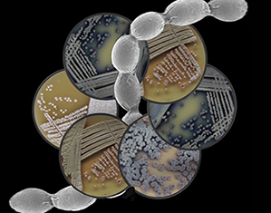
Over the last century, Streptomyces bacteria – and their metabolic products – have revolutionized modern medicine. These little pharmaceutical factories produce a vast array of natural products that have been co-opted for medical and agricultural therapies. In addition to their metabolic sophistication, Streptomyces also exhibit remarkable developmental and regulatory complexity.
Guest-edited by Dr Marie Elliot, this collection of keynote research articles will highlight fascinating aspects of Streptomyces biology, and the advances that are providing us with newfound insight and appreciation for these extraordinary bacteria.
Collection Contents
-
-
Extrusion of extracellular membrane vesicles from hyphal tips of Streptomyces venezuelae coupled to cell-wall stress
More LessExtracellular vesicle release is a wide-spread and broadly important phenomenon in bacteria. However, not much is known about the mechanism of vesicle release in Gram-positive bacteria. Observations of polarly growing Streptomyces venezuelae by live cell time-lapse imaging reveal release of extracellular membrane vesicles from tips of vegetative hyphae. Vesicle extrusion is associated with spontaneous growth arrests, but often the apical cell survives and can re-initiate growth by forming new hyphal branches. Treatment with vancomycin to block peptidoglycan synthesis leads to a high frequency of lysis and vesicle extrusion, where some hyphae can survive growth arrest and vesicle extrusion and reinitiate growth after antibiotic is washed away. The extruded vesicles do not contain nucleoids and do not appear able to proliferate. Vesicle extrusion is not affected by the Ser/Thr protein kinase AfsK that phosphorylates the DivIVA at hyphal tips, nor is it affected by the intermediate filament-like protein FilP that localizes in gradient-like structures at hyphal tips. Notably, hyphae of a scy mutant, which has an unstable apical polarisome structure, are prone to spontaneous growth arrests and vesicle extrusion even in the absence of antibiotic treatment, supporting the idea that the nature of the growth zone at the hyphal tips is important for this route of extracellular vesicle formation. We speculate that the propensity for vesicle extrusion is a direct consequence of how polar growth is organized at hyphal tips in Streptomyces , with the cell-wall sacculus being weak and susceptible to bursting at the apical zones of growth where peptidoglycan synthesis is primarily taking place.
-
-
-
The enigmatic lack of glucose utilization in Streptomyces clavuligerus is due to inefficient expression of the glucose permease gene
More LessStreptomyces clavuligerus ATCC 27064 is unable to use glucose but has genes for a glucose permease (glcP) and a glucose kinase (glkA). Transformation of S. clavuligerus 27064 with the Streptomyces coelicolor glcP1 gene with its own promoter results in a strain able to grow on glucose. The glcP gene of S. clavuligerus encodes a 475 amino acid glucose permease with 12 transmembrane segments. GlcP is a functional protein when expressed from the S. coelicolor glcP1 promoter and complements two different glucose transport-negative Escherichia coli mutants. Transcription studies indicate that the glcP promoter is very weak and does not allow growth on glucose. These results suggest that S. clavuligerus initially contained a functional glucose permease gene, like most other Streptomyces species, and lost the expression of this gene by adaptation to glucose-poor habitats.
-
-
-
Evolution of the PPM-family protein phosphatases in Streptomyces: duplication of catalytic domain and lateral recruitment of additional sensory domains
More LessOriginally identified from eukaryotes, the Mg2+- or Mn2+-dependent protein phosphatases (PPMs) are a diverse group of enzymes whose members include eukaryotic PP2C and some prokaryotic serine/threonine phosphatases. In a previous study, unexpectedly large numbers of PPMs were identified in two Streptomyces genomes. In this work, a phylogenetic analysis was performed with all the PPMs available from a wide variety of microbial sources to determine the evolutionary origin of the Streptomyces PPM proteins. Consistent with earlier hypotheses, the results suggested that the microbial PPMs were relatively recent additions from eukaryotic sources. Results also indicated that the Streptomyces PPMs were divided into two major subfamilies at an early stage of their emergence in Streptomyces genomes. The first subfamily, which contains only six Streptomyces PPMs, possesses a catalytic domain whose sequence and architecture are similar to that of eukaryotic PPMs; the second subfamily contains 89 Streptomyces PPMs that lack the 5a and 5b catalytic domain motifs, similar to the PPMs SpoIIE and RsbU of Bacillus subtilis. Significant gene duplication was observed for the PPMs in the second subfamily. In addition, more than half (54 %) of the Streptomyces PPMs from the second subfamily were found to have at least one additional sensory domain, most commonly the PAS or the GAF domain. Phylogenetic analysis showed that these domains tended to be clustered according to the putative physiological functions rather than taxonomic relationship, implying that they might have arisen as a result of domain recruitment in a late evolutionary stage. This study provides an insight into how Streptomyces spp. may have expanded their PPM-based signal transduction networks to enable them to respond to a greater range of environmental changes.
-
-
-
Effect of Serine Hydroxamate and Methyl α-d-glucopyranoside Treatment on Nucleoside Polyphosphate Pools, RNA and Protein Accumulation in Streptomyces hygroscopicus
More LessThe accumulation of RNA and protein and the kinetics of nucleoside triphosphate and guanosine polyphosphate pools during amino acid starvation and carbon source downshift were investigated in Streptomyces hygroscopicus. RNA accumulation was controlled stringently during both amino acid starvation and carbon source downshift. The pool size of ppGpp increased dramatically under these conditions. However, the intracellular concentrations of nucleoside triphosphates were low and the concentration of guanosine polyphosphates was much lower than in Escherichia coli. The possible significance of this phenomenon in the regulation is discussed.
-