 -
Volume 69,
Issue 12,
2020
Volume 69, Issue 12, 2020
- Antimicrobial Resistance
-
-
-
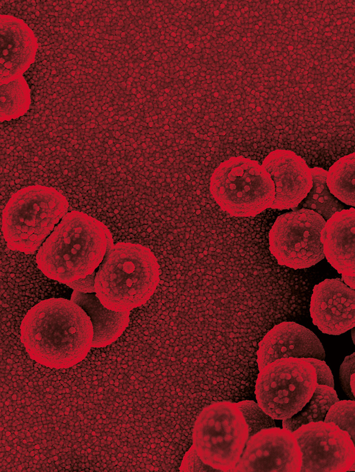
Molecular characterization of methicillin-resistant and -susceptible Staphylococcus aureus recovered from hospital personnel
More LessIntroduction. Methicillin-resistant Staphylococcus aureus (MRSA) is one of the major causes of hospital-acquired infections. Over the past two decades MRSA has become ‘epidemic’ in many hospitals worldwide. However, little is known about the genetic background of S. aureus recovered from hospital personnel in China.
Hypothesis/Gap Statement. The diversity of S. aureus genotypes warrants further surveillance and genomic studies to better understand the relatedness of these bacteria to those recovered from patients and the community.
Aim. The aim of this study was to determine the genetic diversity of MRSA and methicillin-susceptible S. aureus (MSSA) recovered from hospital personnel in Tianjin, North China.
Methodology. Three hundred and sixty-eight hand or nasal swabs were collected from 276 hospital personnel in 4 tertiary hospitals in Tianjin, North China between November 2017 and March 2019. In total, 535 Gram-positive bacteria were isolated, of which 59 were identified as S. aureus . Staphylococcal cassette chromosome mec (SCCmec) typing, multi-locus sequence typing (MLST) and spa typing were performed to determine the molecular characteristics of S. aureus .
Results. Thirty-one out of 276 (11 %) hospital personnel were S. aureus carriers, whereas 11/276 (4 %) carried MRSA. Fifty out of 59 (85 %) S. aureus isolates were resistant or intermediately resistant to erythromycin. The dominant genotypes of MRSA recovered from hospital personnel were ST398-t034-SCCmecIV/V and ST630-t084/t2196, whereas the major genotypes of MSSA included ST15-t078/t084/t346/t796/t8862/t8945/t11653 and ST398-t189/t034/t078/t084/t14014.
Conclusion. Although the predominant genotypes of MRSA recovered from hospital personnel in this study were different from the main genotypes that have previously been reported to cause infections in Tianjin and in other geographical areas of China, the MRSA ST398-t034 genotype has previously been reported to be associated with livestock globally. The dominant MSSA genotypes recovered from hospital personnel were consistent with the those previously reported to have been recovered from the clinic.
-
-
-
-
A novel peptide nucleic acid- and loop-mediated isothermal amplification assay for the detection of mutations in the 23S rRNA gene of Treponema pallidum
More LessIntroduction. Macrolides could be a potential alternative treatment for Treponema pallidum infections in patients; however, macrolide-resistant T. pallidum is spreading rapidly worldwide.
Hypothesis/Gap Statement. There are presently no alternatives to serological tests for syphilis that can be used to evaluate therapeutic effects due to the fact that T. pallidum cannot be cultured in vitro.
Aim. In this study, we constructed a method for rapidly identifying T. pallidum and confirming macrolide resistance by using loop-mediated isothermal amplification (LAMP) with peptide nucleic acids (PNAs).
Methodology. A set of LAMP primers was designed to span nucleotide positions 2058 and 2059 in 23S rRNA. A PNA clamping probe was also designed to be complementary to the wild-type sequence (A2058/A2059) and positioned to interfere with both the annealing of the 3′ end of the backward inner primer and the concomitant extension. Prior to the LAMP assay, swab samples from suspected syphilitic lesions were boiled for DNA extraction.
Results. The assay had an equivalent detection limit of 1.0×101 copies/reaction and showed specificity against 38 pathogens. In the presence of a 4 µM PNA probe, LAMP amplified up to 1.0×101 copies/reaction using plasmids harbouring the complementary mutant sequences (A2058G or A2059G), whereas amplification was completely blocked for the wild-type sequence up to a concentration of 1.0×10³ copies/reaction. For the 66 PCR-positive clinical specimens, the overall detection rate via LAMP was 93.9 % (62/66). Amplification was successful for all 53 mutant samples and was incomplete for all nine WT samples by the PNA-mediated LAMP assays.
Conclusion. We developed a PNA-mediated LAMP method that enabled us to rapidly identify T. pallidum and determine its macrolide susceptibility via a culture-independent protocol.
-
- Clinical Microbiology
-
-
-
Evaluation of GENECUBE Mycoplasma for the detection of macrolide-resistant Mycoplasma pneumoniae
More LessIntroduction. Resistance against macrolide antibiotics in Mycoplasma pneumoniae is becoming non-negligible in terms of both appropriate therapy and diagnostic stewardship. Molecular methods have attractive features for the identification of Mycoplasma pneumoniae as well as its resistance-associated mutations of 23S ribosomal RNA (rRNA).
Hypothesis/Gap Statement. The automated molecular diagnostic sytem can identify macrolide-resistant M. pneumoniae .
Aim. To assess the performance of an automated molecular diagnostic system, GENECUBE Mycoplasma, in the detection of macrolide resistance-associated mutations.
Methodology. To evaluate whether the system can distinguish mutant from wild-type 23S rRNA, synthetic oligonucleotides mimicking known mutations (high-level macrolide resistance, mutation in positions 2063 and 2064; low-level macrolide resistance, mutation in position 2067) were assayed. To evaluate clinical oropharyngeal samples, purified nucleic acids were obtained from M. pneumoniae -positive samples by using the GENECUBE system from nine hospitals. After confirmation by re-evaluation of M. pneumoniae positivity, Sanger-based sequencing of 23S rRNA and mutant typing using GENECUBE Mycoplasma were performed.
Results. The system reproducibly identified all synthetic oligonucleotides associated with high-level macrolide resistance. Detection errors were only observed for A2067G (in 2 of the 10 measurements). The point mutation in 23S rRNA was detected in 67 (26.9 %) of 249 confirmed M. pneumoniae -positive clinical samples. The mutations at positions 2063, 2064 and 2617 were observed in 65 (97.0 %), 2 (3.0 %) and 0 (0.0 %) of the 67 samples, respectively. The mutations at positions 2063 and 2064 were A2063G and A2064G, respectively. The results from mutant typing using GENECUBE Mycoplasma were in full agreement with the results from sequence-based typing.
Conclusion. GENECUBE Mycoplasma is a reliable test for the identification of clinically significant macrolide-resistant M. pneumoniae .
-
-
-
-
Maraviroc, celastrol and azelastine alter Chlamydia trachomatis development in HeLa cells
More LessIntroduction . Chlamydia trachomatis (Ct) is an obligate intracellular bacterium, causing a range of diseases in humans. Interactions between chlamydiae and antibiotics have been extensively studied in the past.
Hypothesis/Gap statement: Chlamydial interactions with non-antibiotic drugs have received less attention and warrant further investigations. We hypothesized that selected cytokine inhibitors would alter Ct growth characteristics in HeLa cells.
Aim. To investigate potential interactions between selected cytokine inhibitors and Ct development in vitro.
Methodology. The CCR5 receptor antagonist maraviroc (Mara; clinically used as HIV treatment), the triterpenoid celastrol (Cel; used in traditional Chinese medicine) and the histamine H1 receptor antagonist azelastine (Az; clinically used to treat allergic rhinitis and conjunctivitis) were used in a genital in vitro model of Ct serovar E infecting human adenocarcinoma cells (HeLa).
Results. Initial analyses revealed no cytotoxicity of Mara up to 20 µM, Cel up to 1 µM and Az up to 20 µM. Mara exposure (1, 5, 10 and 20 µM) elicited a reduction of chlamydial inclusion numbers, while 10 µM reduced chlamydial infectivity. Cel 1 µM, as well as 10 and 20 µM Az, reduced chlamydial inclusion size, number and infectivity. Morphological immunofluorescence and ultrastructural analysis indicated that exposure to 20 µM Az disrupted chlamydial inclusion structure. Immunofluorescence evaluation of Cel-incubated inclusions showed reduced inclusion sizes whilst Mara incubation had no effect on inclusion morphology. Recovery assays demonstrated incomplete recovery of chlamydial infectivity and formation of structures resembling typical chlamydial inclusions upon Az removal.
Conclusion. These observations indicate that distinct mechanisms might be involved in potential interactions of the drugs evaluated herein and highlight the need for continued investigation of the interaction of commonly used drugs with Chlamydia and its host.
-
- Disease, Diagnosis and Diagnostics
-
-
-
Efficient differentiation of Nocardia farcinica, Nocardia cyriacigeorgica and Nocardia beijingensis by high-resolution melting analysis using a novel locus
More LessAccurate identification of Nocardia species remains a challenge due to the complexities of taxonomy and insufficient discriminatory power of traditional techniques. We report the development of a molecular technique that utilizes real-time PCR-based high-resolution melting (HRM) analysis for differentiation of the most common Nocardia species. Based on a novel fusA-tuf intergenic region sequence, Nocardia farcinica , Nocardia cyriacigeorgica and Nocardia beijingensis were clearly distinguished from one another by HRM analysis. The limit of detection of the HRM assay for purified Nocardia spp. DNA was at least 10 fg. No false positives were observed for specificity testing of 20 non-target clinical samples. In comparison to established matrix-assisted laser desorption/ionization-time of flight MS, the HRM assay improved the identification of N. beijingensis . Additionally, all the products of PCR were verified by direct sequencing. In conclusion, the developed molecular assay allows simultaneous detection and differentiation of N. farcinica , N. cyriacigeorgica and N. beijingensis with high sensitivity and specificity.
-
-
-
-
Rapid identification of bacteria directly from positive blood cultures by a modified method using a serum separator tube and matrix-assisted laser desorption ionization – time of flight MS
More LessIntroduction. Several studies have used matrix-assisted laser desorption ionization-time of flight MS (MALDI-TOF) with a serum separator tube (SST) to perform rapid identification of microorganisms directly from positive blood cultures (BCs), with different performances and methodologies.
Hypothesis / Gap Statement. The use of TSS could significantly reduce the time of identification of microorganisms that produce bacteremia.
Aim. Our goals were to evaluate bacterial identification by MALDI-TOF using a method based on an SST and compare it with MALDI-TOF after subculture for 18–24 h.
Methodology. BCs no more than 1 h after a positive growth signal were included in the study. Analysis of results was expressed as a score. Information about time to a positive signal and number of microorganisms was collected.
Results. In total, 253 BCs were analysed; 45.5 % gave a reliable result, 23.3 % an unreliable result and 31.2 % an error in identification. In gram-negative and gram-positive bacteria, the percentages of reliable results were 83.5 and 21.8 %, respectively. According to time to positive signal, the percentages of correct identification and mean score were 81.1 % (99/122) and 1.89±0.30 in Group 1 (<15 h); and 57.2 % (75/131) and 1.70±0.32 in Group 2 (>15 h), respectively (P <0.001). According to the number of microorganisms, the corresponding percentages of correct identification and mean scores were: Group 1 [≤50 microorganisms observed per field (MOF)], 50/94 (53.19 %) and 1.72±0.32; Group 2 (51–100 MOF): 44/66 (66.67 %) and 1.85±0.34; Group 3 (>100 MOF): 79/93 (84.94 %) and 1.84±0.31.
Conclusion. This method allowed us to obtain a high percentage of the aetiological agent of bacteraemia in less than 30 min after a positive BC.
-
- Molecular and Microbial Epidemiology
-
-
-
Seroprevalence of parechovirus A1, A3 and A4 antibodies in Yamagata, Japan, between 1976 and 2017
More LessIntroduction. Although new parechovirus A (PeVA) types, including parechovirus A3 (PeVA3) and PeVA4, have been reported in this century, there have not yet been any seroepidemiological studies on PeVA over a period of several decades.
Hypothesis/Gap Statement. The authors hypothesize that PeVA3 and PeVA4 emerged recently.
Aims. The aim was to clarify changes in the seroprevalence of PeVA1, PeVA3 and PeVA4.
Methodology. Neutralizing antibodies (NT Abs) were measured among residents in Yamagata, Japan in 1976, 1983, 1985, 1990, 1999 and 2017.
Results. The total NT Ab-positive rate for PeVA1 was between 90.7 and 100 % for all years analysed, with that for PeVA3 increasing from 39.6 % in 1976 to 69.6 % in 2017, and that for PeVA4 decreasing from 93.9 % in 1976 to 49.1 % in 2017. The distribution of NT Ab titres for PeVA1, PeVA3 and PeVA4 among those aged less than 20 years old was as follows: those ≥1 : 32 for PeVA1 were between 68.0–89.2 % for all years analysed; those ≥1 : 32 for PeVA3 was 15.4 % in 1976, 44.3–54.9 % in 1983–1990 and 64.8–68.0 % in 1999–2017; and those ≥1 : 32 for PeVA4 were between 49.1–67.2 % in 1976–1990, 41.3 % in 1999 and 23.8 % in 2017.
Conclusions. Our findings in this seroepidemiological study over four decades suggested that PeVA1 has been stably endemic, while PeVA3 appeared around 1970s and has spread since then as an emerging disease, and occasional PeVA4 infections were common in 1970s and 1980s but have been decreasing for several decades in our community.
-
-
-
-
High mortality by nosocomial infections caused by carbapenem-resistant P. aeruginosa in a referral hospital in Brazil: facing the perfect storm
More LessIntroduction. Carbapenem-resistant Pseudomonas aeruginosa is responsible for increased patient mortality.
Gap Statement. Five and 30 day in-hospital all-cause mortality in patients with P. aeruginosa infections were assessed, followed by evaluations concerning potential correlations between the type III secretion system (TTSS) genotype and the production of metallo-β-lactamase (MBL).
Methodology. This assessment comprised a retrospective cohort study including consecutive patients with carbapenem-resistant infections hospitalized in Brazil from January 2009 to June 2019. PCR analyses were performed to determine the presence of TTSS-encoding genes and MBL genes.
Results. The 30-day and 5-day mortality rates for 262 patients were 36.6 and 17.9 %, respectively. The unadjusted survival probabilities for up to 5 days were 70.55 % for patients presenting exoU-positive isolates and 86 % for those presenting exo-negative isolates. The use of urinary catheters, as well as the presence of comorbidity conditions, secondary bacteremia related to the respiratory tract, were independently associated with death at 5 and 30 days. The exoS gene was detected in 64.8 % of the isolates, the presence of the exoT and exoY genes varied and exoU genes occurred in 19.3 % of the isolates. The exoU genotype was significantly more frequent among multiresistant strains. MBL genes were not detected in 92 % of the isolates.
Conclusions. Inappropriate therapy is a crucial factor regarding the worse prognosis among patients with infections caused by multiresistant P. aeruginosa , especially those who died within 5 days of diagnosis, regardless of the genotype associated with TTSS virulence.
-
- Method
-
-
-
Rapid Sepsityper in clinical routine: 2 years’ successful experience
More LessIntroduction. Rapid identification of the causative agent of sepsis is crucial for patient outcomes.
Aim. The Sepsityper sample preparation method enables direct microbial identification of positive blood culture samples via matrix-assisted laser desorption ionization/time-of-flight mass spectrometry (MALDI-TOF MS).
Hypothesis/Gap statement. The implementation of the Sepsityper method in the routine practice could represent a fundamental tool to achieve a prompt identification of the causative agent of bloodstream infections, and therefore accelerate the adoption of the proper antibiotic treatment.
Methodology. In this study, the novel rapid workflow of the MALDI Biotypr Sepsityper kit (Bruker Daltonik GmbH, Germany) was evaluated using routine samples from a 2-year period (n=6918), and dedicated optimized protocols for the microbial groups that were more difficult to identify were developed. Moreover, the use of the residual bacterial pellet to perform susceptibility testing using different methods (commercial broth microdilution, disc diffusion, gradient diffusion) was investigated.
Results. The rapid Sepsityper protocol allowed the identification of 5470/6338 (86.3 %) monomicrobial samples at species level, with very good performance for all of the clinically most significant pathogens (2510/2592 enterobacteria, 631/669 Staphylococcus aureus and 223/246 enterococci were identified). Streptococcus pneumoniae , Bacteroides fragilis and yeasts were the most troublesome to identify, but the application of specific optimized protocols significantly improved their rate of identification (from 14.7–71.5 %, 47.8–89.7 % and 37.1–89.5 %, respectively). Specificity was 100 % (no identification was made for the false-positive samples). Further, the residual pellet proved to be suitable to investigate susceptibility to antimicrobials, enabling us to simplify the workflow and shorten the time to report.
Conclusion. The Rapid Sepsityper workflow proved to be a reliable sample preparation method for identification and susceptibility testing directly from positive blood cultures, providing novel approaches for accelerated diagnostics of bloodstream infections.
-
-
Volumes and issues
-
Volume 73 (2024)
-
Volume 72 (2023 - 2024)
-
Volume 71 (2022)
-
Volume 70 (2021)
-
Volume 69 (2020)
-
Volume 68 (2019)
-
Volume 67 (2018)
-
Volume 66 (2017)
-
Volume 65 (2016)
-
Volume 64 (2015)
-
Volume 63 (2014)
-
Volume 62 (2013)
-
Volume 61 (2012)
-
Volume 60 (2011)
-
Volume 59 (2010)
-
Volume 58 (2009)
-
Volume 57 (2008)
-
Volume 56 (2007)
-
Volume 55 (2006)
-
Volume 54 (2005)
-
Volume 53 (2004)
-
Volume 52 (2003)
-
Volume 51 (2002)
-
Volume 50 (2001)
-
Volume 49 (2000)
-
Volume 48 (1999)
-
Volume 47 (1998)
-
Volume 46 (1997)
-
Volume 45 (1996)
-
Volume 44 (1996)
-
Volume 43 (1995)
-
Volume 42 (1995)
-
Volume 41 (1994)
-
Volume 40 (1994)
-
Volume 39 (1993)
-
Volume 38 (1993)
-
Volume 37 (1992)
-
Volume 36 (1992)
-
Volume 35 (1991)
-
Volume 34 (1991)
-
Volume 33 (1990)
-
Volume 32 (1990)
-
Volume 31 (1990)
-
Volume 30 (1989)
-
Volume 29 (1989)
-
Volume 28 (1989)
-
Volume 27 (1988)
-
Volume 26 (1988)
-
Volume 25 (1988)
-
Volume 24 (1987)
-
Volume 23 (1987)
-
Volume 22 (1986)
-
Volume 21 (1986)
-
Volume 20 (1985)
-
Volume 19 (1985)
-
Volume 18 (1984)
-
Volume 17 (1984)
-
Volume 16 (1983)
-
Volume 15 (1982)
-
Volume 14 (1981)
-
Volume 13 (1980)
-
Volume 12 (1979)
-
Volume 11 (1978)
-
Volume 10 (1977)
-
Volume 9 (1976)
-
Volume 8 (1975)
-
Volume 7 (1974)
-
Volume 6 (1973)
-
Volume 5 (1972)
-
Volume 4 (1971)
-
Volume 3 (1970)
-
Volume 2 (1969)
-
Volume 1 (1968)
Most Read This Month


